ULTRASOUND

Ultrasound - GE Voluson Signature Series S-8

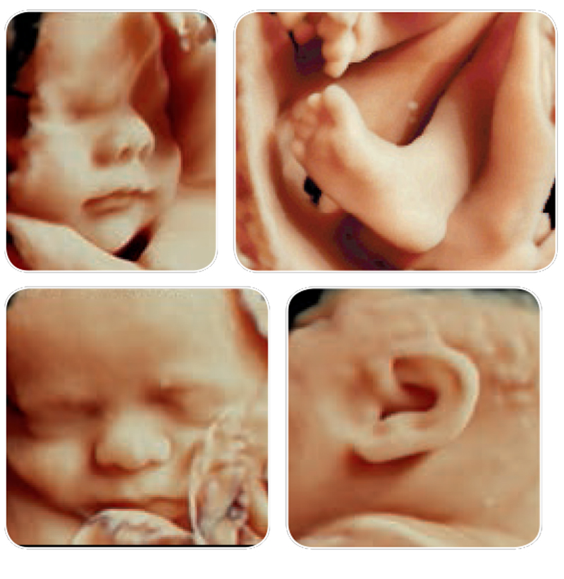
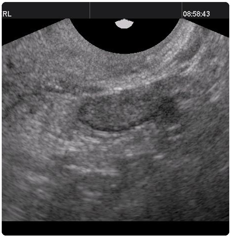
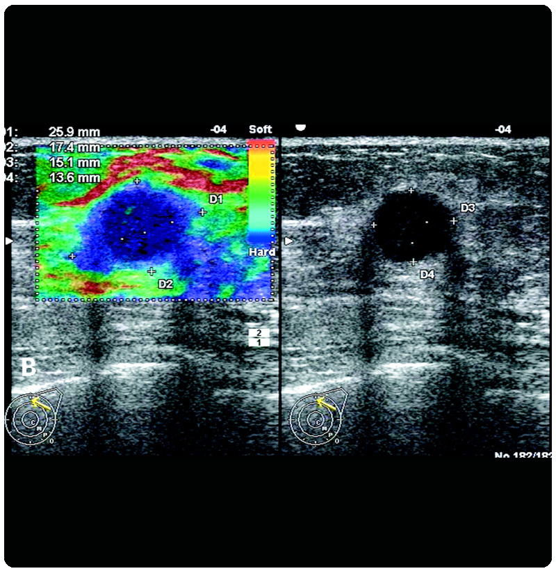
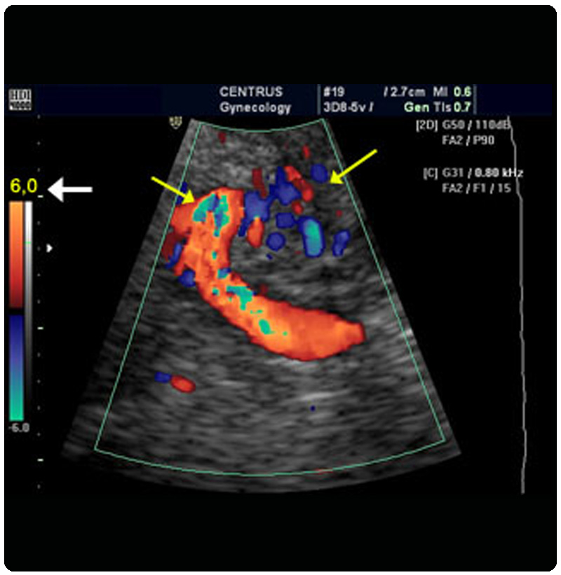
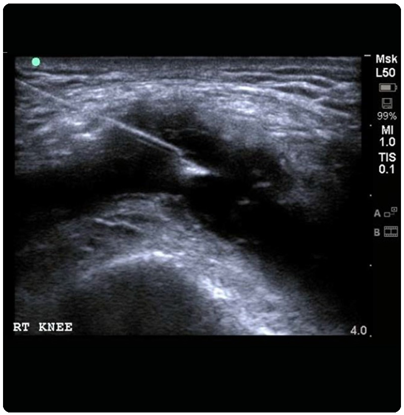
Ultrasound imaging uses sound waves to produce pictures of the inside of the body. It is used to help diagnose the causes of pain, swelling and infection in the body’s internal organs and to examine a baby in pregnant women and the brain and hips in infants. It’s also used to help guide biopsies, diagnose heart conditions, and assess damage after a heart attack. Ultrasound is safe, noninvasive, and does not use ionizing radiation.


  • Innovative probe technology giving Extraordinary image quality and diagnostic confidence.
  • Sophisticated fetal assessment and women imaging.
  • 3D/ 4D imaging with Omniview& TUI (Tomographic Ultrasound Imaging).
  • HD live giving realistic skin effects and exceptional anatomical realism.
  • Advanced color Doppler and power Doppler for ultra slow flows.

...


Clinical Applications

Obstetrics and Fetal imaging:

  • Early viability and dating scan (1st 10 weeks) with 3D imaging of yolk sac and embryo.
  • 1st trimester NT scan for nuchal translucency and intracranial translucency.
  • Level II scan at 18-20 weeks for complete anomaly assessment including fetal heart, spine, brain, limbs, thorax, abdominal organs &soft markers for aneuploidy.
  • Fetal well being and growth scan (20 weeks till term).
  • Doppler study of MCA, umbilical artery, uterine arteries, ductus venosus and aorta for early diagnosis of IUGR, uteroplacental insufficiency, fetal hypoxia and fetal anemia in Rh incompatible pregnancy, twin-twin transfusion, etc.
  • 3D and 4D fetal ultrasound for fetal morphology, face visualization, cleft lip/palate, congenital malformations, skeletal dysplasia, spina bifida/meningocele, dwarfism, club feet, fetal heart 4D, fetal movements and gestures, reactivity/tonicity to rule out neuromyopathy, placenta evaluation for any abnormalities like lower segment insertion, etc.
  • Introducing shortly USG guided Fetal interventions – Amniocentesis, Chorionic villus sampling.

...


Gynaecology & Infertility imaging:

  • Various ovarian, uterine and cervical pathologies detection, characterization and extent visualization.
  • 3D imaging of uterus for uterine shape anomalies, intrauterine adhesions, endometrial polyps, submucosal fibroids, IUCD localization, adnexal imaging for hydrosalpinx.
  • TUI Volume imaging gives the exact volume measurement of endometrial hyperplasia, cysts, polyps, fibroids, tumors. Virtual hysteroscopy using the slicing technique.
  • Follicular monitoring – AutomaticSonoAVC follicle and 3D multiplanar ovarian imaging gives enhanced measurement consistency and increased patient comfortability by reducing exam time.

...


Small parts:

  • Breast- Extended view for better visualization.
  • Breast elastography for lesion characterization.
  • Thyroid imaging with HD flow.
  • Scrotum and testicular imaging.
  • Soft tissue tumor characterization and extent.

...


Colour Doppler imaging:

  • Upper and lower limbs arterial Doppler for plaques; stenosis in PVD and embolic detection.
  • Upper and lower limbs venous Doppler for superficial; deep venous thrombosis.
  • Varicose veins evaluation and perforator detection.
  • Carotid and vertebral vessels Doppler for CAD patients, stroke patients with auto IMT measurement; plaque detection.
  • Splenoportal axis for cirrhosis, portal hypertension and thrombosis.
  • Abdominal aorta and iliac vessels for aneurysms/dissection.
  • Renal Doppler for renal artery stenosis, hypertension; post-transplant patients.

...


USG guided Interventions:

  • USG guided FNAC; biopsy.
  • Aspiration; drainage of abscess, collection, pleural effusion, ascites, etc.
  • Shortly introducing - Fetal interventions (Amniocentesis; CVS).

...

Ultrasound services in Himachal

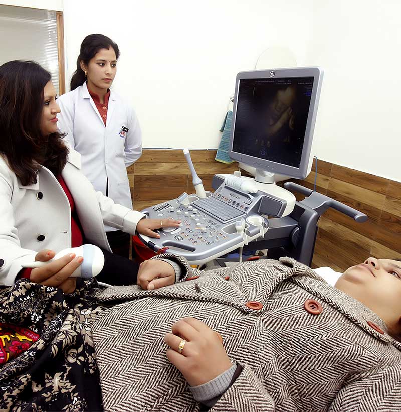
Apex diagnostics offering an extensive range of Ultrasound services in Sirmour, Solan and Shimla, In order to enquire or know more please visit our website.

our partners

Pre Insurance Health Check Partner.

Refer a Patient

We are committed to improving people’s lives through personalized health care. When you refer your patient to us, we are pleased to assist you with the diagnosis, treatment and monitoring of your patients’ care

find us